
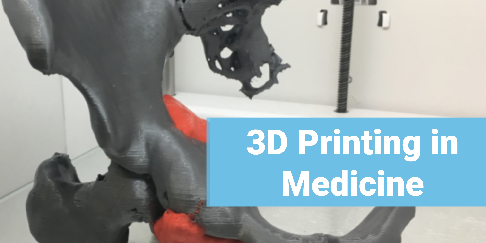
Bitfab collaborated with one of the most advanced 3D printing projects in medicine in Spain: the 3D printing laboratory of the Gregorio Marañón Hospital in Madrid, now the Advanced Planning and 3D Manufacturing Unit (UPAM3D). This article, written in 2019, explains how the laboratory worked and what kind of cases were solved there.
The credit for the project goes to Rubén and Jose, the doctors who created it, whom we talk about below.
The variety of medical 3D printing projects done there is enormous: biomodels and guides made directly for the doctors, who then use them to communicate, operate, try out new techniques… all in real clinical cases, helping real patients.
📖 What do you want to read?
What is the Gregorio Marañón’s FabLab (now UPAM3D)?
The Marañón FabLab is a 3D printing laboratory integrated into the Hospital, where doctors come every day to solve their healthcare problems with 3D printing. Healthcare means directly helping patients with completely customized models, although research, simulation and teaching models… are also made for innovative projects within the Hospital.
Who has developed it?
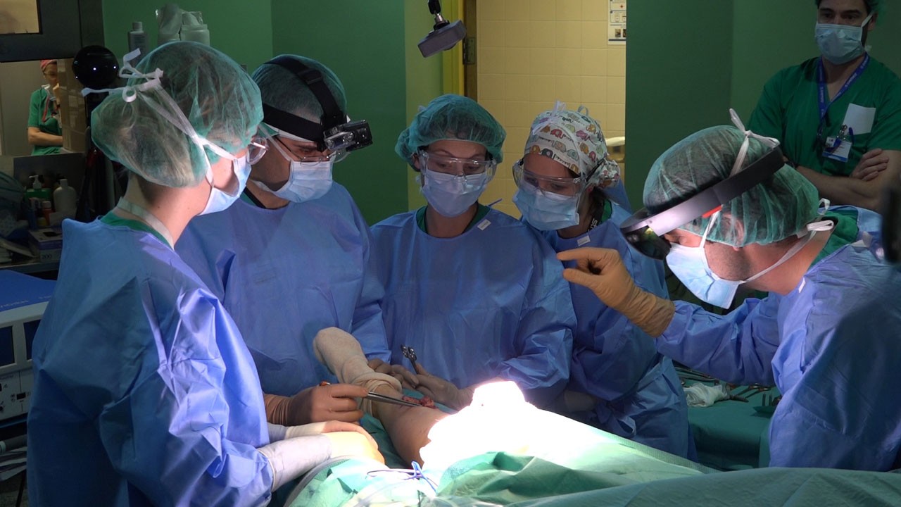
The Marañón FabLab is a project founded by Rubén Pérez-Mañanes and Jose Antonio Calvo Haro and has been operating for many years. They are two oncology trauma surgeons who saw the possibilities of 3D desktop printing within a hospital and started the project with their own means.
You can find them and follow them on social media:
Other engineers have worked in the lab, such as Guillermo Rodríguez, who presented the project in this talk at T3chFest 2017 (in Spanish), as well as undergraduate and graduate students from different universities in Madrid who train there in biomedical 3D printing.
The workflow of the Gregorio Marañón Hospital
How is the typical case printed in the Hospital’s laboratory? This was the workflow and the software used in each phase. You will see many free and open tools and desktop printers: the laboratory’s philosophy is open, and it aims to make 3D printing in medicine much more accessible with desktop solutions within everyone’s reach:
- Communication. Usually the doctor (especially surgeons of all specialties) goes to the laboratory with the case he wants to print, what he needs it for, what kind of product he wants to print (biomodels, surgical guides, splints, instruments, research projects…). It is an unparalleled opportunity to be in direct contact with the doctors, which is the advantage of having the 3D printing laboratory integrated directly into the Hospital.
- Medical imaging. The starting point of the cases is the medical image in DICOM format: a CT scan, an MRI, etc.
- Segmentation. This is where the medical image, which comes in layers, is turned into a 3D file that can be printed, modified, visualized, etc. The tools used are 3D Slicer (open and free), Horos (open and free) or proprietary solutions such as Philips Advanced Visualization Workspace (formerly IntelliSpace Portal).
- 3D modeling. The meshes generated by the segmentation programs cannot always be printed directly. It is often necessary to process them to improve printability or to modify them. For this, Meshmixer, free software from Autodesk, is used. It is essential for surgical guides: with it, cutting templates are created so that the surgeons cut exactly where they have previously planned in 3D.
- Slicing. Parts are sliced with Cura by UltiMaker and PreForm for the resin prints.
- Printing. At the time, the laboratory had professional desktop printers, simple but powerful machines that make it possible to produce parts every day at a reasonable cost: Ultimaker (2, 3 and S5), Witbox 2 and Form 2 from Formlabs for the resin parts.
- Publication of all cases. The FabLab philosophy is open, so all cases are shared on Twitter and Linkedin. You can see it yourself in the lab’s twitter account.
What is done in the laboratory? Examples of 3D printing applications in medicine
Biomodels
Biomodels are 3D printed parts from image segmentation, anatomical models of the patient’s body parts to be visualized or physically held.
What are biomodels for? They fulfill the following functions:
- Communication between doctors who plan treatments and surgeries
- Communication with patients
- Surgical planning, trials… Checking if the instruments can access the area to be operated on, choosing a prosthesis size before operating, pre-folding plates before putting them on the patient, etc.
Simulation and teaching
A very interesting case of simulation is the model made for Dr. Javier Río: a complete aorta printed in TPU (flexible) and PVA (soluble) so that he could try a new surgical technique.
Also in teaching, Dr. Juan de León designs models of hearts for a course in diagnosing heart disease in fetuses by means of ultrasound scans.
Surgical guides
Surgical guides are templates to guide cuts and punctures during surgery. They are made in biocompatible materials with resin printers and are used mainly in interventions to remove tumors, because they allow planning the operation in 3D in advance and cutting with precision to remove the least amount of healthy tissue possible.
Navigation and Augmented Reality
One of the fields where the Marañón Hospital is a pioneer together with the Carlos III University is in navigation operations and augmented reality. Anatomical markers (that fit in a fixed position on the patient) and cameras located around the operating room are used to guide the surgeon in 3D during surgery for complex cases such as resection (extraction) of pelvic tumors:
Thank you very much for reading 🙂